November 9, 2016
MRI
An MRI (Magnetic Resonance Imaging) scan is typically the preferred test for people who may have a brain tumor. It shows a better picture of the brain than a CT scan, although a CT scan may be the first test that is done.
After the initial testing, more diagnostic tests may be needed. If the MRI shows a possible primary brain tumor (a brain tumor that originates in the brain), patients normally do not need other imaging tests of the body.
The patient must lie still inside the MRI machine for about an hour while it generates images of the inside of the brain. During the scan, three different views are typically taken of the head and neck to show a tumor’s location and position. Using these views, different scans can be done to produce exact images that best show the tumor and the area around the brain.
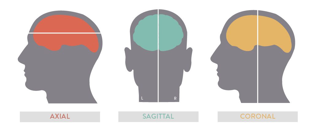
Axial: This is a straight line passing through a spherical body between two poles and that the body may revolve around
Sagittal: This is a vertical plane passing through the standing body from front to back
Coronal: This is a vertical plane from head to foot that is parallel to the shoulders
After the images are taken, the medical provider or radiologist will read the digital images and create a report which your doctor will review the results with you.